
The Knee – A Masterpiece of Biomechanics
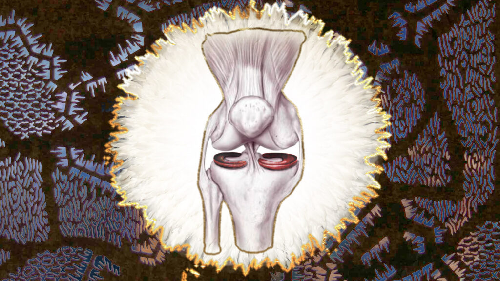
The knee is the largest joint in the human body and is subjected to enormous stress every day. With every step, it supports the body’s weight, absorbs shock, and provides both stability and mobility. This constant mechanical load, however, also makes the knee particularly vulnerable to injury and wear.
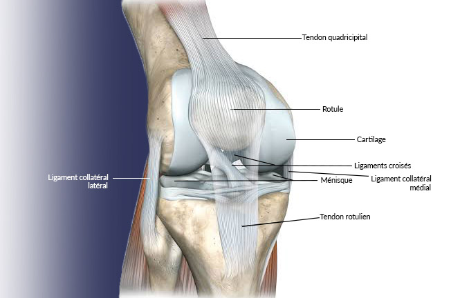
Its complex anatomy allows all movements to work together with remarkable precision. The knee joint connects the thigh bone (femur) to the shin bone (tibia). This interaction is supported by the menisci, cruciate ligaments, collateral ligaments, cartilage, tendons and muscles, all of which work together to ensure stability and an even distribution of forces across the joint.
Meniscal Injuries
The two menisci are crescent-shaped cartilage discs located between the femur and tibia. They increase the contact surface within the knee joint, act as natural shock absorbers and distribute pressure evenly across the joint cartilage. At the same time, they play a crucial role in stabilising the knee.
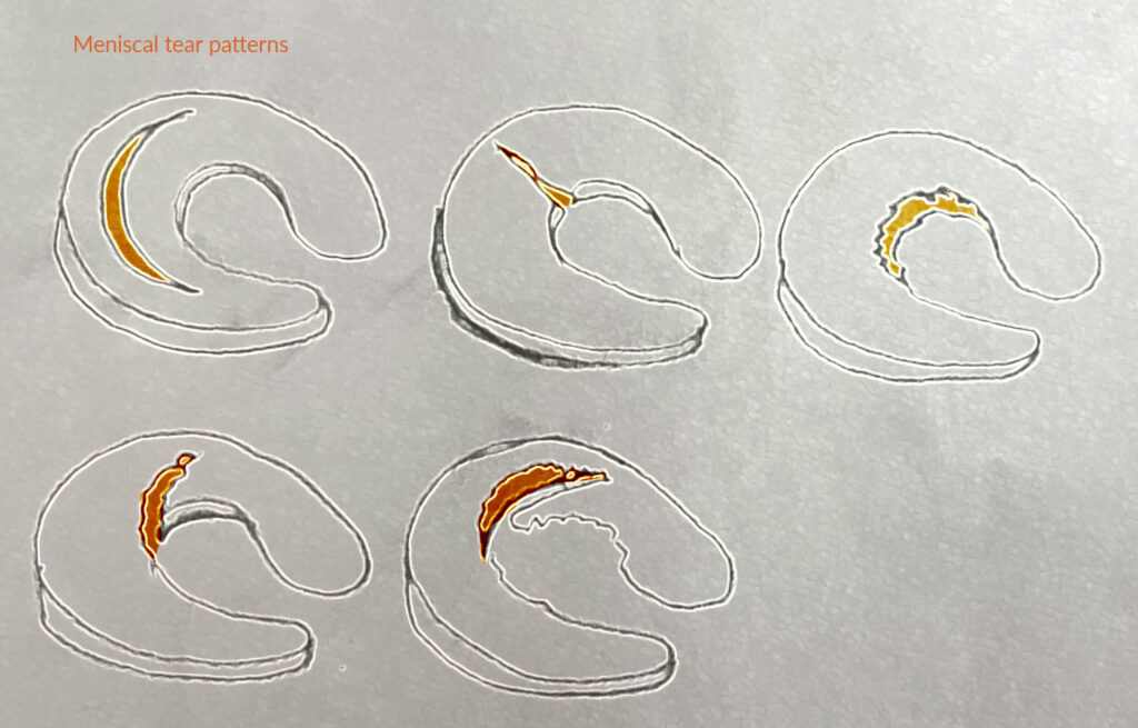
Meniscal injuries may occur either as a result of an acute trauma, such as a sports injury, or through age-related degeneration. Smaller tears can often be treated conservatively with rest, ice therapy, anti-inflammatory medication and targeted physiotherapy. More extensive tears may require arthroscopic surgery. Whenever possible, the damaged meniscus is repaired by suturing the tear. If this is not feasible, only the damaged portion is removed (partial meniscectomy) in order to preserve as much healthy tissue as possible and reduce the risk of developing osteoarthritis later in life.
Cruciate Ligament Injuries
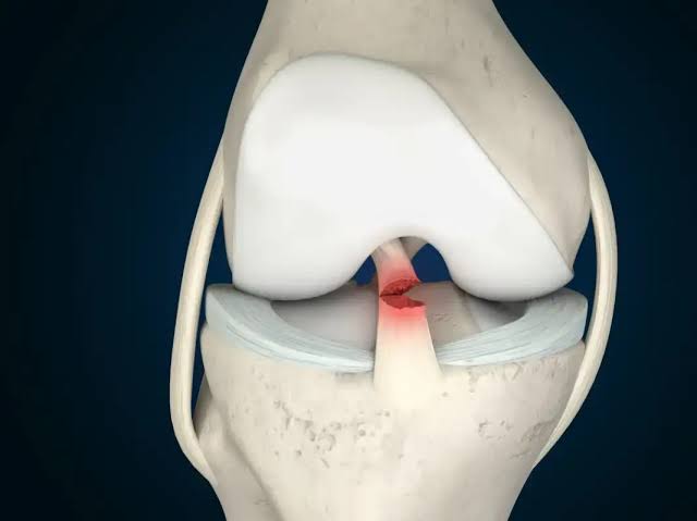
The anterior and posterior cruciate ligaments connect the femur and tibia. Crossing each other in the centre of the knee joint, they prevent excessive forward and backward movement of the bones, thereby providing essential stability, particularly during rapid changes of direction.
A cruciate ligament tear usually results from a sudden twisting motion or excessive stretching of the knee, most commonly during sporting activities. Meniscal or collateral ligament injuries frequently occur at the same time. Typical symptoms include severe pain, swelling, instability and restricted movement.
Depending on the severity of the injury, as well as the patient’s age and activity level, treatment may involve conservative management or arthroscopically assisted reconstructive surgery.
Baker’s Cyst
A Baker’s cyst is a fluid-filled swelling of the joint capsule located at the back of the knee. It is not a disease in itself but usually develops as a consequence of another knee condition, such as a meniscal tear, osteoarthritis or chronic inflammation.
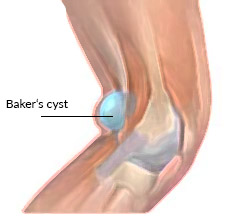
When the knee becomes irritated or inflamed, it produces excess synovial fluid. As pressure within the joint increases, the joint capsule may bulge backwards, resulting in the formation of a Baker’s cyst.
Small Baker’s cysts often cause no symptoms and may disappear once the underlying condition has been treated. Larger cysts can cause a feeling of tightness behind the knee, pain or restricted movement. In rare cases, numbness or swelling of the calf may occur if nearby nerves or blood vessels become compressed.
Patellar Tendinopathy (Jumper’s Knee)
The kneecap (patella) is a small bone embedded within the tendon of the quadriceps muscle. Although it is not technically part of the knee joint itself, it plays a vital role in transmitting the force generated by the knee’s extensor mechanism.
Patellar tendinopathy, commonly known as Jumper’s Knee, is characterised by painful irritation of the patellar tendon where it attaches to the lower pole of the kneecap. It most commonly affects athletes involved in sports requiring frequent jumping or rapid changes of direction, such as volleyball, basketball and athletics.
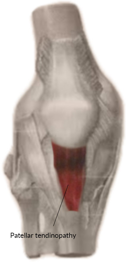
In addition to repetitive sporting activities, factors such as leg malalignment, foot deformities, tight muscles, congenital ligament laxity and age-related changes in tendon structure can increase the risk of developing this condition.
Typical symptoms include activity-related pain just below the kneecap, particularly when jumping, climbing stairs or after prolonged sitting.
Knee Osteoarthritis (Gonarthrosis)
Knee osteoarthritis, also known as gonarthrosis, is the progressive degeneration of the articular cartilage within the knee joint. As cartilage gradually wears away, the joint surfaces rub more directly against one another, leading to pain, stiffness and reduced mobility.
Typical symptoms include stiffness and pain when taking the first steps after resting, pain while climbing stairs and a gradual decline in the knee’s ability to bear weight. Factors such as ageing, excess body weight, leg malalignment and previous injuries all increase the risk of developing knee osteoarthritis.
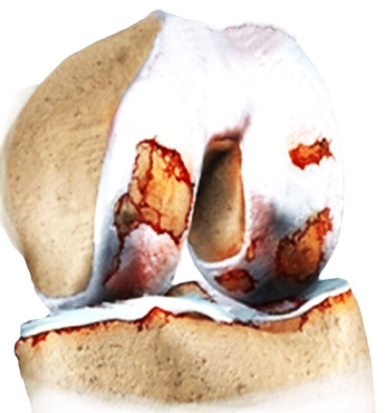
Typical symptoms include stiffness and pain when taking the first steps after resting, pain while climbing stairs and a gradual decline in the knee’s ability to bear weight. Factors such as ageing, excess body weight, leg malalignment and previous injuries all increase the risk of developing knee osteoarthritis.
Because cartilage has only a limited capacity to regenerate, early treatment focuses on slowing disease progression, relieving symptoms and preserving joint function for as long as possible.
Knee Bursitis
Bursae are small fluid-filled sacs that act as cushions between bones, tendons and the skin, reducing friction during movement. When one of these bursae becomes inflamed, the condition is known as bursitis.
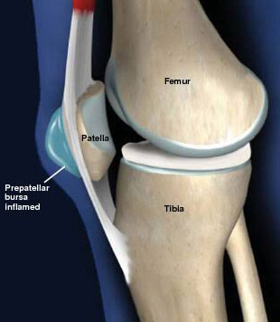
Inflammation of the bursa located in front of the kneecap (prepatellar bursitis) typically presents with noticeable swelling at the front of the knee. The affected area is often red, warm and tender to the touch.
Common causes include repeated kneeling, chronic pressure on the knee or minor injuries. Less commonly, bursitis may be caused by a bacterial infection. Metabolic disorders such as gout or diabetes, as well as inflammatory rheumatic diseases, can also increase the likelihood of developing bursitis.
Modern Treatment Options for Knee Disorders
Treatment is always tailored to the underlying cause, the extent of the damage and the individual needs of each patient. The primary goals are to relieve pain, restore mobility and, whenever possible, preserve the natural knee joint over the long term.
In many cases, conservative treatment is the first choice. This may include targeted physiotherapy, muscle strengthening exercises, anti-inflammatory medication, knee braces or orthoses and, where appropriate, weight reduction. If leg malalignment contributes to the problem, customised orthopaedic insoles or corrective realignment procedures may also be beneficial.
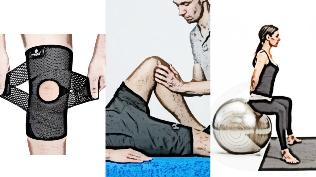
Postural training methods that focus on improving body alignment and muscular balance may also complement conservative treatment in selected patients. The CANTIENICA® Method for Body in Evolution, for example, aims to optimise functional alignment through activation of the deep postural muscles and may support body awareness and movement quality. For international patients staying in Vienna, an introduction to the method can be combined with their rehabilitation programme. As online sessions are available in English, patients can continue practising after returning home as part of a long-term approach to postural control and functional movement.
Innovative regenerative therapies, including stem cell treatment, may help stimulate the body’s natural healing processes and promote the regeneration of damaged tissue in selected patients. These therapies are particularly promising for cartilage defects and early stages of osteoarthritis and may help delay the need for joint replacement surgery.
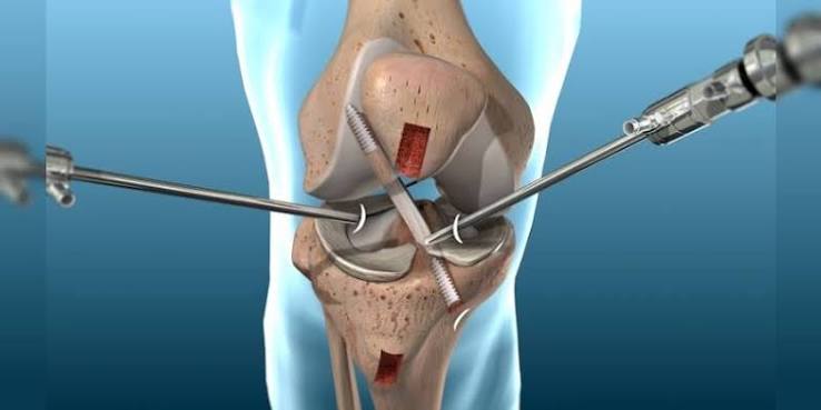
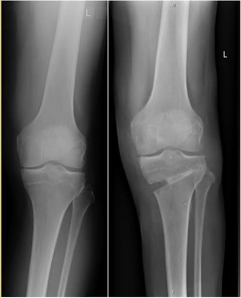
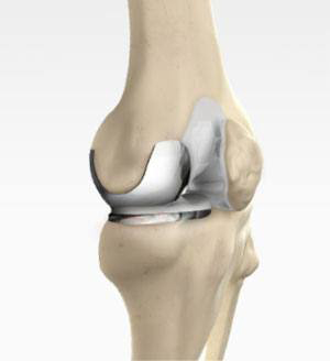
When conservative treatment no longer provides sufficient relief, a range of minimally invasive surgical procedures is available. These include arthroscopic meniscal and cruciate ligament surgery, joint-preserving corrective osteotomy and, in cases of advanced osteoarthritis, partial or total knee replacement.
When Should You See a Knee Specialist?
Persistent knee pain lasting more than a few days, recurring symptoms or restricted mobility should always be assessed by an orthopaedic specialist. Swelling, feelings of instability, locking of the knee joint or pain following an injury are also warning signs that require medical evaluation. An early diagnosis often allows for joint-preserving treatment and can help prevent long-term damage.
Sources: Gesundheitsinformation.de; arthrose-behandlung.at; orthinform.de; Schünke M et. al., Prometheus: Lernatlas der Anatomie (Allgemeine Anatomie und Bewegungssystem) Thieme, 5. Auflage; Aumüller G et al., Duale Reihe Anatomie, Thieme, 5. Auflage; Isabel Haider-Strutz Anatomie, Biologie, Physiologie für Gesundheitsberufe 3. Auflage; https://stiwell.medel.com/orthopaedics/gonarthrosis-(knee-osteoarthritis); stiwell.medel.com