
Next-Generation Sequencing – Some Background
Next-generation sequencing (NGS) is a modern technique used to sequence and analyse DNA and RNA in parallel with the help of computational tools. In oncology, NGS is used, among other things, to investigate the molecular characteristics of a tumour in as much detail as possible.
By analysing the tumour genome, genetic alterations can be identified that may play a role in tumour growth and development. This information can help improve the understanding of a tumour’s biological characteristics and identify specific alterations that may be relevant to treatment.
NGS therefore provides an important foundation for precision oncology. Treatment decisions are based not only on the organ in which a tumour originated, but increasingly also on the individual molecular alterations present in the tumour cells.
What is the Genome?
The genome is the complete set of genetic information of an organism. Put simply, it can be thought of as the body’s biological blueprint.
In humans, this information is contained in somatic cells, or body cells, in 23 pairs of chromosomes, making a total of 46 chromosomes. Germ cells, by contrast, namely egg cells and sperm cells, contain only a single set of 23 individual chromosomes. They are therefore haploid, whereas body cells are diploid.
Chromosomes consist of DNA (deoxyribonucleic acid), which is closely associated with proteins, particularly histones. DNA contains numerous genes. Simplified, a gene is a specific segment of DNA located at a defined position, known as a gene locus, that carries the information needed to produce a functional product, such as a protein or a functional RNA molecule.
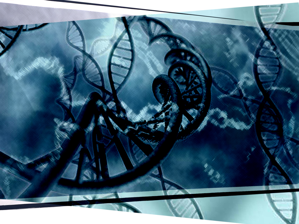
Chemically, DNA consists of four different organic bases: adenine (A), thymine (T), guanine (G) and cytosine (C). Each base is attached to a sugar molecule, deoxyribose, which gives DNA its name, and a phosphate group. Together, these three components form a nucleotide, the basic building block of DNA.
DNA predominantly exists as a double strand in the form of a double helix. The two strands are connected by complementary base pairs: adenine pairs with thymine (A–T), while guanine pairs with cytosine (G–C). These base pairs are held together by hydrogen bonds. Among other functions, this complementary structure enables the cell to replicate genetic information reliably.
A typical protein-coding gene contains several functional regions. These include regulatory regions such as the promoter, which plays an important role in initiating transcription, as well as a transcribed region containing exons and, in many genes, introns. At the end of transcription, termination signals ensure that RNA polymerase stops the transcription process. Exons are the segments that remain in the mature mRNA after RNA processing. In protein-coding genes, they contain the coding sequences relevant to the production of the protein. Introns, by contrast, are removed from the pre-mRNA during the so called splicing process.
The size of genes varies considerably. Some genes comprise only a few hundred nucleotides, while others span millions of bases. The human genome contains approximately 20,000 protein-coding genes. In addition, there are numerous genes that produce functional, non-protein-coding RNAs. The exact number therefore also depends on how a gene is defined and which types of genes are included.
In humans, DNA is located predominantely in the cell nucleus. A small proportion of genetic informaton is also found in the mitochondria.
Sources:
https://www.genome.gov/genetics-glossary/Genome
https://www.genome.gov/about-genomics/fact-sheets/Deoxyribonucleic-Acid-Fact-Sheet
From DNA to RNA
RNA (ribonucleic acid) plays a central role in the transcription of genetic information. Like DNA, RNA consists of nucleotides. However, its surgar molecule is ribose, and instead of the base thymine (T), RNA contains uracil (U). The four bases of RNA are therefore adenine (A), uracil (U), guanine (G), and cytosine (C).
During transcription, the information contained in a segment of DNA is transferred into an RNA sequence by the enzyme RNA polymerase. In the case of a protein-coding gene, the initial product is a molecule known as pre-mRNA, which still contains both exons and introns.
The pre-mRNA is subsequently processed through several steps to produce mature mRNA. These include splicing, during which the introns are removed and the remaining exons are joined together, as well as the addition of a 5′ cap structure and a poly (A) tail. Among other fuctions, thes modifications contribute to mRNA stability and facilitate its further processing and use within the cell.
The mature mRNA can then leave the nucleus and enter the cytoplasm. There, itsgenetic information is translated into a protein at the ribosomes. This process is known as translation.
DNA and RNA differ not only in their chemical composition. Usually, DNA exists predominantly as a stable double helix and primarly serves as the long-term storage medium for genetic information. RNA, on the contrary, is single-stranded and can adopt a wide variety of structures and perform different functions, depending on its role. mRNA serves as a temporary copy of the informtion contained in a gene and provides the template for the production of specific proteins.
Sources:
https://www.ncbi.nlm.nih.gov/books/NBK26887
NGS in Modern Oncology

Today, it is well established that cancer arises from changes in the genetic material. These changes can affect genes that regulate cell growth and division. Certain genetic alterations can cause cells to multiply uncontrollably, leading to a formation of a tumour.
NGS can be used to analyse both the DNA and RNA of a tumour. This usually involves examining a tumour tissue sample. The aim is to characterize the molecular alteration of the tumour as precisely as possible and identify which of these alterations may play a role in its growth.
This has contributed to the development of precision oncology. Rather than focusing exclusively on the type of cancer and the organ affected, clinicians can increasingly take into account the individual molecular characteristics of each tumour. The results of NGS analysis can help guide treatment towards a more targeted approach based on the biological properties of the specific tumour.
Sources:
https://pmc.ncbi.nlm.nih.gov/articles/PMC9588890
https://www.cancer.gov/ccg/research/genome-sequencing
https://www.cancer.gov/ccg/research/cancer-genomics-overview
https://www.esmo.org/insights-by-esmo
What Tumour DNA Reveals
The analysis of tumour DNA provides information about genetic alterations present in the genetic material of tumour cells. In simplified terms, these can be described as “permanent errors in the blueprint.”
This means that the DNA sequence of the tumour cell has changed. When such an alteration is present in a tumour cell, it is generally passed on to daughter cells during subsequent cell divisions. As a result, the alteration can spread throughout the tumour.
However, not every genetic alteration identified is necessarily responsible for the development or growth of the tumour. Molecular analysis therefore places particular importance on distinguishing between alterations that are merely incidental and those known as driver alterations, which promote tumour growth.
NGS can detect various types of genetic alterations.

1. Point Mutations
A point mutation involves a change in a single base in the DNA sequence. For example, at a specific position, one base (A, T, G, or C) may be replaced by another base.
Such a change can alter the information used to produce the corresponding protein. If this results in a different amino acid being incorporated into the protein, its structure or function may change. However, not every base substitution automatically leads to a change in the protein.
A well-known example is the BRAF V600E mutation, which occurs, among other cancers, in melanoma (black skin cancer). At position 600 of the BRAF protein, the amino acid valine (V) is replaced by glutamic acid (E). This alteration can cause the BRAF protein to remain permanently activated, continously transmitting growth signals within the cells.
Such a molecular alteration can represent an important point of attack for targeted therapy. The aim is to specifically inhibit the signalling pathway that has becomee overactive as a result of the mutation.
Sources:
https://pubmed.ncbi.nlm.nih.gov/26825657
2. Insertions and Deletions
An insertion occurs when one or more bases are added to the DNA sequence. A deletion occurs when one or more bases are lost.
Depending on the size and position of the alteration, it can significantly affect the function of the gene involved. For example, if bases are inserted or deleted and their number is not a multiple of three, the entire reading frame of a gene can shift. This is known as a frameshift mutation.
The result may be a substantially altered or prematurely truncated protein that can no longer perform its original function.
One example is deletions in exon 19 of the EGFR gene, which occur in certain types of lung cancer. These alterations in the DNA sequence also change the structure of the EGFR protein. In some cases, this can cause the receptor to become permanently activated and transmit growth signals to the cell.
Such alterations can also be relevant for targeted therapy, as the resulting overactivity of the EGFR signalling pathway can be specifically inhibited.
Sources:
https://pubmed.ncbi.nlm.nih.gov/35151115
3. Amplifications – Gene Multiplications
In an amplification, a specific segment of DNA is present in a significantly increased number of copies. The DNA sequence of the gene itself does not necessarily change. Rather, more copies of the gene or gene segment are present than would normally be expected.
The increased copy number can, under certain circumstances, cause the cell to produce substantially more RNA and, consequently, more of the corresponding protein.
A well-known example is HER2 amplification in certain types of breast, gastric, and oesophageal cancer. The increased number of HER2 gene copies can lead to a substantial increase in the amount of HER2 protein on the surface of tumours cells. As a result, HER2 can transmit stronger growth signals and contribute to tumour development.
When such an amplification is detectd, it can provide important information for treatment decisions. In this case, treatments can be used that specifically target the protein that is present in increased amounts or is overactive.
Sources:
https://pubmed.ncbi.nlm.nih.gov/21505336
Why RNA Sequencing Can Make Gene Fusions Easier to Detect
Analysis of tumour RNA can often make certain gene fusions easier to detect than analysis at the DNA level. In a gene fusion, DNA segments that originally belong to different genes become joined together as a result of a chromosomal rearrangement. The breakpoints are often located within introns, which are non-protein-coding regions of a gene. These regions are removed during the formation of mature RNA through a process known as splicing.
In RNA, by contrast, the resulting fusion transcript can be detected directly: the coding regions of the two originally seperate genes appear joined together. This often allows sequencing software to identify a gene fusion more directly.
Although the underlying alteration originates at the DNA level, detecting it there can be more challenging. One reason is that introns can be very large and structurally complex, extending over many thousands of bases and sometimes containing repetitive sequences. During RNA processing, these regions have already been removed, making the fusion-relevant connection between the coding sequences much easier to identify.
Sources:
https://pmc.ncbi.nlm.nih.gov/articles/PMC6859817
https://pmc.ncbi.nlm.nih.gov/articles/PMC6437215
https://www.cancer.gov/ccg/research/genome-sequencing/cgci/using-cgci/technology
Tissue-Specific or Tumour-Agnostic?
Certain genes are associated with characteristic or recurrent chromosomal breaks and rearrangements. These can repeatedly give rise to the same or similar gene fusions. Some of these gene fusions are closely associated with a particular type of cancer, while others can occur across a wide range of tumour types.
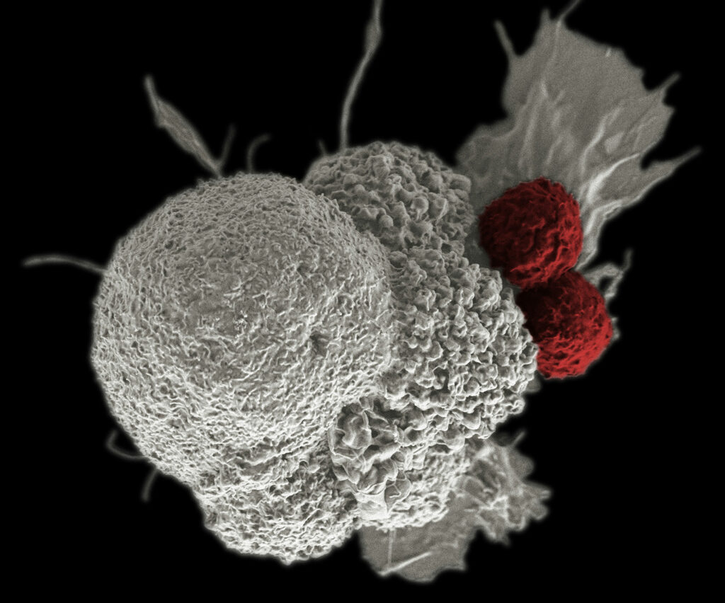
Modern pharmaceutical research has made it possible to develop drugs specificaly designed to block the disease-promoting proteins or signalling pathways produced by such a gene fusions. this makes the gene fusion itself a potential therapeutic target.
To understand their significance in precision medicine, gene fusions can be broadly divided into two groups:
1. Tissue-Specific Gene Fusions
These fusions occur predominantly in a particular type of cancer and can represent a characteristic molecular feature of the disease.
A classic example is the Philadelphia chromosome (BCR::ABL1) in chronic myeloid leukaemia (CML).
This characteristic chromosomal alteration is found in more than 95 per cent of patients with CML. It involves an exchange of genetic material between chromosomes 9 and 22. The ABL1 gene on chromosome 9 becomes joined to the BCR gene on chromosome 22, resulting in the formation of the BCR::ABL1 fusion gene. The resulting fusion protein has a permanently activated tyrosine kinase, which promotes the uncontrolled proliferation of leukaemia cells.
Today, drugs known as tyrosine kinase inhibitors (TKIs) can specifically block the activity of the BCR::ALB1 fusion protein. This targeted approach has fundamentally transformed the prognosis of CML. For most patients with chronic-phase CML, the disease can now be controlled over the long term, and life expectancy can approach that of the general population.
Sources:
https://www.cancer.gov/types/leukemia/hp/cml-treatment-pdq
https://www.cancer.gov/research/progress/discovery/gleevec
2. Tumour-Agnostic Gene Fusions
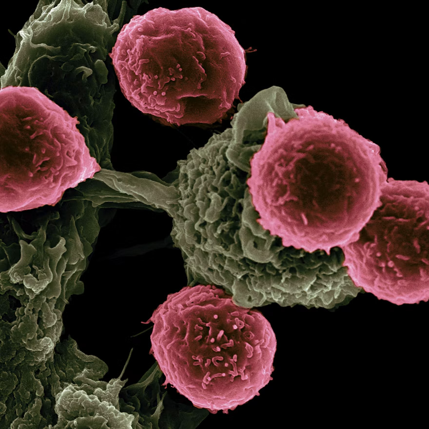
Other gene fusions are not restricted to a particular organ. They can occur in different types of cancer, where they can act as so-called driver alterations. For these tumours, the key question is not simply where the cancer originated, but which molecular alteration is driving its growth.
For certain tumour-agnostic gene fusions, the same targeted therapy can therefore be used across different cancer types. The prerequisite is that the relevant molecular alteration with therapeutic significance has been identified.
A particularly illustrative example is NTRK gene fusions. The NTRK1, NTRK2, and NTRK3 genes can fuse with numerous different partner genes. This fusion can lead to permanent activation of the tyrosine kinase section of the NTRK gene, which plays a key role in tumour development. In simplified terms, this permanently switches on a molecular growth signal within the cell.
NTRK fusions can occur in many tumour types, including lung, thyroid, and colorectal cancers. However, they are rare in most of these common cancer types.
For this very reason, it is important to identify such genetic alterations specifically. If an NTRK gene fusion is detected, targeted treatment with a TRK inhibitor may be an option under certain circumstances, regardless of the organ in which the tumour originated. These drugs target the dysregulated activity of the TRK protein resulting from the gene fusion. The key factor in treatment selection is therefore not solely the type of tumour, but also which molecular alterations have been identified in the tumour cells and whether these findings provide a basis for a specific therapeutic option.
This changes the way to view cancer: the question “Where did the tumour originate?” is no longer the sole determining factor in treatment. Instead, another question becomes equally important: “Which molecular alteration is driving the tumour, and is there a drug that can target it specifically?”
Sources:
https://pmc.ncbi.nlm.nih.gov/articles/PMC7903287
https://pubmed.ncbi.nlm.nih.gov/42090631
https://www.onkopedia.com/de/onkopedia/guidelines/praezisionsonkologie/@@guideline/html/index.html
What Happens During NGS
In simplified terms, an NGS workflow can be divided into four main steps. The exact procedure may vary depending on the sequencing technology used and whether DNA or RNA is being analysed.
1. Fragmentation
In a DNA-based NGS analysis, the DNA is first broken down into smaller fragments. This can be achieved, for example, using enzymes, ultrasound (sonication), or transposase-based methods. A transposase is an enzyme encoded by a transposon-related gene.
In an RNA-based analysis, RNA is firstl isolated from the tumour tissue. Because RNA is less stable than DNA and cannot be analysed directly by all sequencing platforms, it is generally first converted into complementary DNA (cDNA). This cDNA can then be used to prepare the sequencing library.
2. Adapter Ligation and Preparation of the DNA Library
Short, specially designed DNA sequences known as adapters are attached to the DNA or cDNA fragments.
These adapters enable the fragments to bind to the sequencing instrument and facilitate subsequent processing.
The complete set of DNA or cDNA fragments prepared in this way are known as the sequencing library.
3. Amplification and Sequencing
Depending on the technology used, the fragments are amplified to generate enough copies for the subsequent measurement. In certain sequencing methods, this process produces so-called DNA clusters, each consisting of many identical copies of a DNA fragment.
The sequences of the fragments are then determined by identifying the order of the four DNA building blocks: adenine (A), cytosine (C), guanice (G), thymine (T). In an RNA-based analysis, the cDNA derived from the RNA is sequenced accordingly.
The sequencing platform initially generates measurement signals, which the software then uses to determine the corresponding base sequences.
4. Bioinformatic Data Analysis
Sequencing generates a large amount of data, which is subsequently processed and analysed using bioinformatics. For example, the sequences obtained are compared with a reference sequence of the human genome to identify genetic alterations.
Depending on the testing method used, tumour NGS can detect mutations, gene fusions, changes in gene copy number, and other relevant molecular alterations.
The alterations identified can then be assessed in terms of their biological significance and potential relevance for treatment.
Sources:
https://pmc.ncbi.nlm.nih.gov/articles/PMC4351865
https://pmc.ncbi.nlm.nih.gov/articles/PMC9291972
https://pmc.ncbi.nlm.nih.gov/articles/PMC6476432
Why NGS Is Useful
Traditional diagnostic methods such as immunohistochemistry (IHC), fluorescence in situ hybridisation (FISH), and polymerase chain reaction (PCR) are designed to detect specific molecular alterations or biological characteristics. NGS (Next-Generation Sequencing), by contrast, makes it possible to analyse numerous genes and different types of genetic alterations simultaneously.
This comprehensive analysis can help tailor treatment more precisely to the individual molecular characteristics of a tumour.
For a long time, cancer was assessed primarily according to the tissue in which the tumour originated and the type of tumour involved. As a result, patients with the same type of cancer often received similar treatments. The individual biological differences between tumours could only be taken into account to a limited extent. For example, a drug might cause a tumour to shrink in Patient A, have no effect on Patient B, and cause significant side effects in Patient C.
Today, NGS represents an important step towards personalised precision oncology. Two people with, for example, lung cancer may have tumours that differ considerably at the molecular level.
Depending on the test, hundreds of cancer-related genes can be analysed simultaneously. The results provide the treating physician with a kind of molecular “fingerprint” of the tumour. Such alterations can provide important indications as to whether a particular targeted therapy may be appropriate .

Together with the type of cancer, the tumour stage, other diagnostic findings, and the patient’s individual circumstances, this information can be taken into account when determining the most appropriate treatment.
Sources:
https://www.cancer.gov/publications/pdq/information-summaries/genetics/overview-hp-pdq